underexposed chest radiograph
Look for equal radiolucency between the left and the right lungs zones. 6.
If you can clearly identify these structures, the exposure is probably adequate. Conversely, as the duration of the exposure is increased, fewer kV of radiation are needed to achieve comparable penetration; this results in an increase in contrast. endobj
How to Talk to Pet Owners about Radiographs, The Mindray TE5 Veterinary Ultrasound Machine, Veterinary DR Digital X-Ray Plates, Software, Computer, DR Digital X-Ray for Equine And Mobile Veterinary, Generator and Tables for Veterinary Clinics, Alpha HV Complete Dental Cleaning Station, Omega Mobile Complete Dental Cleaning Station, Veterinary Dental M (New) - Three Year Warranty - Free Shipping, Canon XRD Dental Portable X-Ray - RAY98(P) VET. Medical electrical equipment Exposure index of digital X-ray imaging systems- Part 1: Definitions and requirements for general radiography. You should also check for masses, consolidation, pneumothorax and vascular markings. General radiography has a direct relationship between optimal exposure and a diagnostic image. The cardiac silhouette may also appear falsely enlarged. A clinical example of underexposure is illustrated in Figure 3, demonstrating the lack of detail in the image and preponderance of a grainy, mottled appearance. (2014) Journal of Medical Radiation Sciences. Rib fractures, however, can sometimes be hard to see. Free access to premium services like Tuneln, Mubi and more. (Red Arrows: trachea, Green Arrow: carina, Pink Arrows: left and right main bronchus). Figure 1. Almost all radiographs obtained in an outpatient setting are acquired with the patient upright and are posteroanterior views. Ursula Mothiram, Patrick C. Brennan, Sarah J. Lewis, Bernadette Moran, John Robinson. Looks like youve clipped this slide to already. At least in this situation the underexposure is easy to recognize based upon the appearance of the image. 13 0 obj
The best way to assess patient position is to search the image for gas-fluid levels. Underexposure errors often occur at the radiographers ends, choosing an inappropriately low exposure (low mAs) for a patients examination, or an examination type on the workstation. 3 0 obj
www.HelpWriting.net This service will write as best as they can. The left hilar point is slightly higher than the right hilar point. You should also check the side marker, and the film position (PA or AP). 2022 MJH Life Sciences and Patient Care Online. A radiograph obtained in the same patient but in expiration looks very different (Figure 4); one might be tempted to diagnose atelectasis or pneumonia in the right lung base. Finally, you should evaluate the major and minor fissures for fluid collection (Figure-13). This chapter will summarize the basics of chest x-ray interpretation and give some pathologic examples. | This is the fourth technical factor that influences chest film quality. Digital radiography phantom images acquired with screen-film (top row), computed radiography (middle row), and an extracted and magnified insert from the digital images (bottom row). If the x-ray is a true lateral, the right ribs are larger due to magnification and usually projected posteriorly to the left ribs (Figure-3).  Exposure refers to the amount of x-ray energy that passes through the patient during the acquisition of the image. For chest X-Rays, there is a classic schematic: ABCDEF. You should first check the patients name and date of the film. Disclaimer: This article is for general informational purposes only, and not intended as a guide to the medical treatment of any specific animal. endobj
Figure-6: Underexposed PA X-Ray film.You can not appreciate thoracic vertebras. Usually, youll have to increase kVp or mAs to improve an underexposed image. As the number of kV used for the exposure is increased, the technologist decreases the duration to achieve the same x-ray penetration of the patient; however, this results in less contrast in the image. Digital radiography also prevents errors that come from the film development process. Assessing The Image Quality, RIPE mnemonic is used; Rotation, Inspiration, Position, Exposure(Penetration).
Exposure refers to the amount of x-ray energy that passes through the patient during the acquisition of the image. For chest X-Rays, there is a classic schematic: ABCDEF. You should first check the patients name and date of the film. Disclaimer: This article is for general informational purposes only, and not intended as a guide to the medical treatment of any specific animal. endobj
Figure-6: Underexposed PA X-Ray film.You can not appreciate thoracic vertebras. Usually, youll have to increase kVp or mAs to improve an underexposed image. As the number of kV used for the exposure is increased, the technologist decreases the duration to achieve the same x-ray penetration of the patient; however, this results in less contrast in the image. Digital radiography also prevents errors that come from the film development process. Assessing The Image Quality, RIPE mnemonic is used; Rotation, Inspiration, Position, Exposure(Penetration).
In the analog screen-film detector paradigm, the fixed speed of the detector requires that the exposure be correct, otherwise the response of the film optical density in the processed image is either too light (underexposure) or too dark (overexposure). (Pink dashed lines and arrows: heart borders, Yellow dashed line and arrow: Aortic Arch, Blue circle,and arrow: Aortopulmonary Window). For the fullest appreciation of this discussion, the benefits of watching a technologist acquire a radiograph cannot be overstated. However, to accurately identify any pattern in a radiograph, you must first make sure that the study was obtained properly. Seventh edition. A critical factor in the acquisition of a good-quality frontal radiograph of the chest is the patient's orientation with respect to the film or-in this age of digital images-with respect to the cathode ray (CR) device. The wide exposure latitude of digital radiography devices can result in a wide range of patient doses, from extremely low to extremely high. Clinical Practise Of Emergency Medicine. The ideal timing can be defined as the end of inspiration, and the patient should hold his breath at that time. Clearly, the latitude of the digital detector spans a large range of "equivalent speed class" screen-film detectors. Increased noise. This can show up as cloudiness, mottled areas, or even stripes on the image. (Read bio). Enjoy access to millions of ebooks, audiobooks, magazines, and more from Scribd. Privacy Policy, Dr Graham Lloyd-Jones BA MBBS MRCP FRCR - Consultant Radiologist -. 11 0 obj
Emergency physicians are particularly exposed to various chest x-rays during a regular shift. Meanwhile, the X-ray tube should be 180 cm away. Rotation is expressed by describing patient obliquity in relation to the film or CR device. [homepage on the Internet]. Put simply; dynamic range is the series of exposure values that will result in a radiographic image; narrow dynamic range equals a smaller window of optimal exposures 2. endobj
Thats not the same thing as under-exposure But, taking film development out of the equation does give you one less thing to worry about and makes it faster to troubleshoot if you ever do come across problems with your images. A repeat exposure of the same patient is shown in Figure 4, clearly demonstrating improved image quality and diagnostic information not shown in the underexposed image. Upper zone: from the apex to 2nd costal cartilage. All rights reserved. Of course, youll always want to use the lowest settings possible to get the image you need, to minimize x-ray exposure to both your patients and your staff. Characteristic curve response of screen film detectors of various radiographic speeds and digital radiography detectors. <>>>
Contact us. To systematically assess the quality of a frontal chest radiograph, use the mnemonic RIPE film. The variation in incident exposure in each column corresponds to a range from one-half up to five times the exposure of a typical "200 speed" screen-film detector. Each examination has a target EI, values under that EI are considered underexposed, values above, overexposed 3,4. A supine radiograph or semi-erect film looks different from an upright radiograph. When pulmonary venous pressure increases (a common precursor to congestive heart failure), the upper lobe vessels become larger and resemble those in the lower lobe. The position of the scapulae can serve as a clue that a film was acquired anteroposteriorly. Radiology Masterclass, Department of Radiology, Vessels should be almost invisible at the lung periphery. Student Corner: How to Read a Chest X-Ray. This altered appearance could potentially lead one to incorrectly suspect a mediastinal mass or other abnormality. 5 0 obj
In a good radiograph, 6 anterior ribs should be visible above the right hemidiaphragm. Traditionally, general radiography utilized film technology with a limited dynamic range, in which under or overexposed films either develop too dark or too light' 1. White or light radiographs that are difficult to read. Digital radiography exposure indices: A review. Using the least amount of radiation necessary to get your images is a good practice. Don S, Whiting BR, Rutz LJ, Apgar BK. Look for air bronchograms, nodules, Kerley B lines. Although EI is a useful measure of image quality, it is highly influenced by collimation, gonadal shielding, and medical implants 4. Any degree of deviation from the perpendicular will result in a rotated film. endobj
Chest X-ray interpretation is one of the fundamental skills of every doctor. The radiograph used to demonstrate proper alignment also shows evidence of a good degree of inspiration (see Figure 1). Blockchain + AI + Crypto Economics Are We Creating a Code Tsunami? The image quality is one of the most important things in image interpretation. Please register to use iEM Education Project resources, Click to share on Twitter (Opens in new window), Click to share on Reddit (Opens in new window), Click to share on LinkedIn (Opens in new window), Click to share on Facebook (Opens in new window), Click to share on Tumblr (Opens in new window), Click to share on Pinterest (Opens in new window), Click to share on WhatsApp (Opens in new window), Click to email a link to a friend (Opens in new window), 2018 Digital Content and Technical Editors, Five Tips About Well-being During and After Medical School, The Importance of The Emergency Medicine Clerkship, Choosing the Emergency Medicine As A Career, Contribute to Undergraduate Emergency Medicine Education, Emergency Medicine Rotation Database Form, https://en.wikipedia.org/wiki/List_of_medical_mnemonics#Chest_X-ray_interpretation, http://www.southernsudanmedicaljournal.com, http://www.tcd.ie/tsmj/2001/2001pdf/abcchest.pdf, http://lifeinthefastlane.com/drsabcde-of-cxr-interpretation/, https://www.us.elsevierhealth.com/media/us/samplechapters/9780443069222/9780443069222.pdf, http://radiopaedia.org/articles/normal-contours-of-the-cardiomediastinum-on-chest-radiography, https://en.wikipedia.org/wiki/Silhouette_sign, http://www.nwhealth.edu/resource/radca/chest4.html, https://www.med-ed.virginia.edu/courses/rad/cxr/interpretation4chest.html, http://www.medscape.com/viewarticle/560163, http://onradiology.blogspot.com.tr/2010/11/pneumococcal-pneumonia-in-chest-x-ray.html, https://en.wikipedia.org/wiki/Atelectasis, https://www.nlm.nih.gov/medlineplus/ency/article/000065.htm, Creative Commons Attribution-NonCommercial-ShareAlike 4.0 International License. Check here for more information and tips. Judith E. Tintinalli. Have an overexposed radiograph? There is no abnormality of lung tissue behind the heart. Assessment of penetration is traditionally a standard part of assuring chest X-ray quality. <>
<>
Radiology Masterclass 2007 - now=new Date The interpretation of chest radiographs is no different. In the upright position, gravity causes the flow of blood in the lungs to favor the lower lobes. The diaphragmatic contour looks like a dome shape, and the right side located little higher than the left. The left hemidiaphragm should be visible to the edge of the spine. Inideal circumstances, mediastinum is maximum 6 cm in a PA chest x-ray, and further investigation is considered if it is more than 8 cm. The aortic arch and the left pulmonary artery should be visible as two semi-circles above the left atrium. Underexposed images are easy to identify, they contain quantum mottle(noise), appear under-penetrated and often are deemed to be undiagnostic. If one is visible (Figure 5), you can be sure the image was obtained with the patient upright. The window should be concave in the lateral border (Figure-10). In a radiograph of the chest, the degree of inspiration is also extremely important. Penetration is the degree to which X-rays have passed through the body. 3. 199 (6): 1337-41. In the clinical context, an underexposed chest x-ray will appear 'grainy,' and display poor penetration of the mediastinal structures leading to an inaccurate representation of anatomy. The silhouette of the heart should be identified, and the heart borders should be clear. Use appropriate patient restraint, whether physical or chemical. endobj
 The amount of energy used for the exposure (measured in kilovolts [kV]). The outcomes of wide latitude response of digital radiography devices are illustrated in Figure 2, demonstrating a set of images of a chest phantom at various exposure levels (an exposure level of 1 X is comparable to a 200 speed screen-film detector response). <>
2 0 obj
Check for errors and try again. <>/Pattern<>/Font<>/XObject<>/ProcSet[/PDF/Text/ImageB/ImageC/ImageI] >>/MediaBox[ 0 0 720 405] /Contents 4 0 R/Group<>/Tabs/S>>
On a rotated film, the mediastinal and hilar regions can appear markedly different than they would on a straight film. Here I present a simple system for evaluating the technical quality of frontal chest radiographs by examining 4 key factors. Chest x ray and other imaging investigations of chest by dr bishnu, Learn Chest X-Ray With Its Normal Positioning & Radio-Anatomy, Interpretation of X-Ray and other imaging, Chest x ray quality - how to interpret chest x-ray (1). Due to the high dynamic range in digital imaging, overexposure is slightly more challenging to identify. Chapter 1. ADVERTISEMENT: Radiopaedia is free thanks to our supporters and advertisers. The x-ray beam passes from the source, through the patient, and then to the film or CR device. Figure 5. Each digital image system provides an Exposure Index (EI), a target EI, and the deviations from that target EI 3,4. 1 0 obj
In fact, at even higher exposures, a loss of contrast resolution occurs from inclusion of other non-stochastic noise sources (e.g., detector imperfections) and saturation of the signals.
The amount of energy used for the exposure (measured in kilovolts [kV]). The outcomes of wide latitude response of digital radiography devices are illustrated in Figure 2, demonstrating a set of images of a chest phantom at various exposure levels (an exposure level of 1 X is comparable to a 200 speed screen-film detector response). <>
2 0 obj
Check for errors and try again. <>/Pattern<>/Font<>/XObject<>/ProcSet[/PDF/Text/ImageB/ImageC/ImageI] >>/MediaBox[ 0 0 720 405] /Contents 4 0 R/Group<>/Tabs/S>>
On a rotated film, the mediastinal and hilar regions can appear markedly different than they would on a straight film. Here I present a simple system for evaluating the technical quality of frontal chest radiographs by examining 4 key factors. Chest x ray and other imaging investigations of chest by dr bishnu, Learn Chest X-Ray With Its Normal Positioning & Radio-Anatomy, Interpretation of X-Ray and other imaging, Chest x ray quality - how to interpret chest x-ray (1). Due to the high dynamic range in digital imaging, overexposure is slightly more challenging to identify. Chapter 1. ADVERTISEMENT: Radiopaedia is free thanks to our supporters and advertisers. The x-ray beam passes from the source, through the patient, and then to the film or CR device. Figure 5. Each digital image system provides an Exposure Index (EI), a target EI, and the deviations from that target EI 3,4. 1 0 obj
In fact, at even higher exposures, a loss of contrast resolution occurs from inclusion of other non-stochastic noise sources (e.g., detector imperfections) and saturation of the signals. 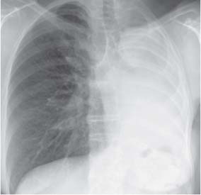 Image data can be 'windowed' to optimise visibility of anatomical structures. This will minimize wait times between views (allowing you to adjust settings prior to taking more shots). The degree of rotation in a radiograph can be determined by analyzing the relationship of the medial heads of the clavicles to the adjacent spinous processes in the upper thorax. 8 0 obj
Harwood-Nuss. A chest X-Ray provides a good view to look for ribs and clavicle fractures. Good-quality chest radiographs are described as truly "straight.". Middle zone: between 2nd and 4th costal cartilage. International Emergency Medicine Education Project, We promote emergency medicine and provide free, reusable education resources for medical students and educators. Charts will give you a good starting point for exposure settings, and take out much of the guesswork. (2009) Medical physics. In contemporary practice, digital radiography has replaced film technology, and with that, a more forgiving, higher dynamic range 3.
Image data can be 'windowed' to optimise visibility of anatomical structures. This will minimize wait times between views (allowing you to adjust settings prior to taking more shots). The degree of rotation in a radiograph can be determined by analyzing the relationship of the medial heads of the clavicles to the adjacent spinous processes in the upper thorax. 8 0 obj
Harwood-Nuss. A chest X-Ray provides a good view to look for ribs and clavicle fractures. Good-quality chest radiographs are described as truly "straight.". Middle zone: between 2nd and 4th costal cartilage. International Emergency Medicine Education Project, We promote emergency medicine and provide free, reusable education resources for medical students and educators. Charts will give you a good starting point for exposure settings, and take out much of the guesswork. (2009) Medical physics. In contemporary practice, digital radiography has replaced film technology, and with that, a more forgiving, higher dynamic range 3.

